New laser imaging technology for real-time monitoring of animal conditions
Release date: 2017-05-26

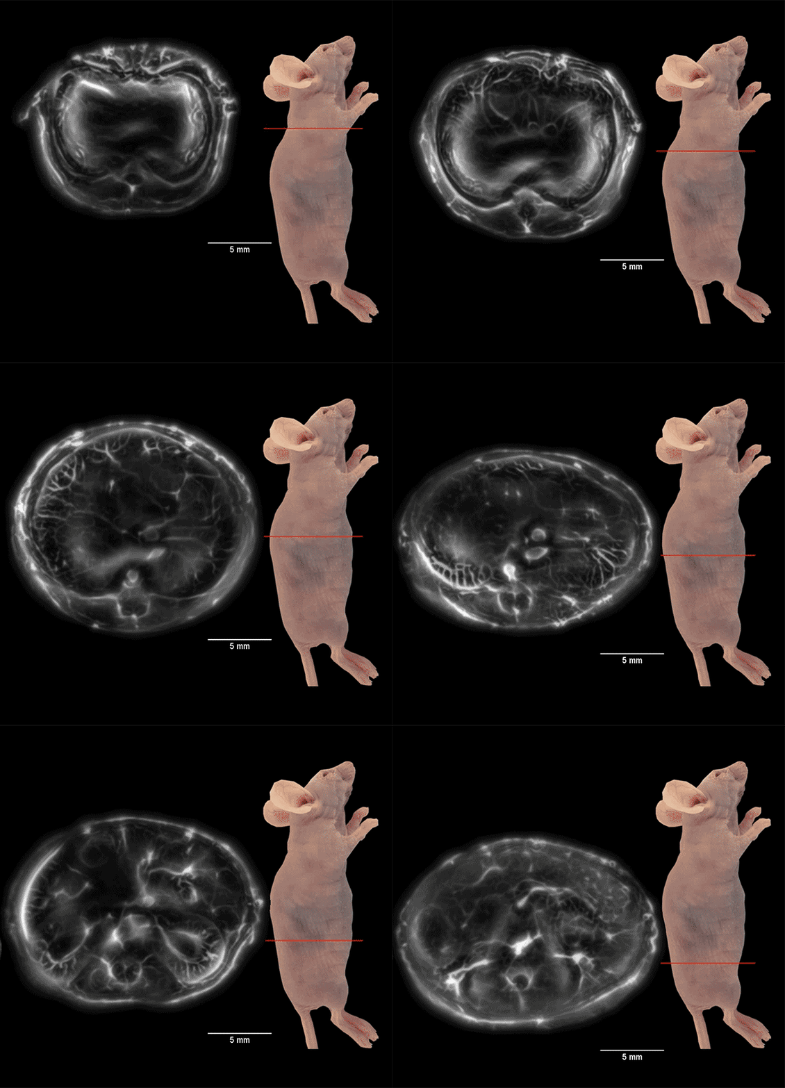
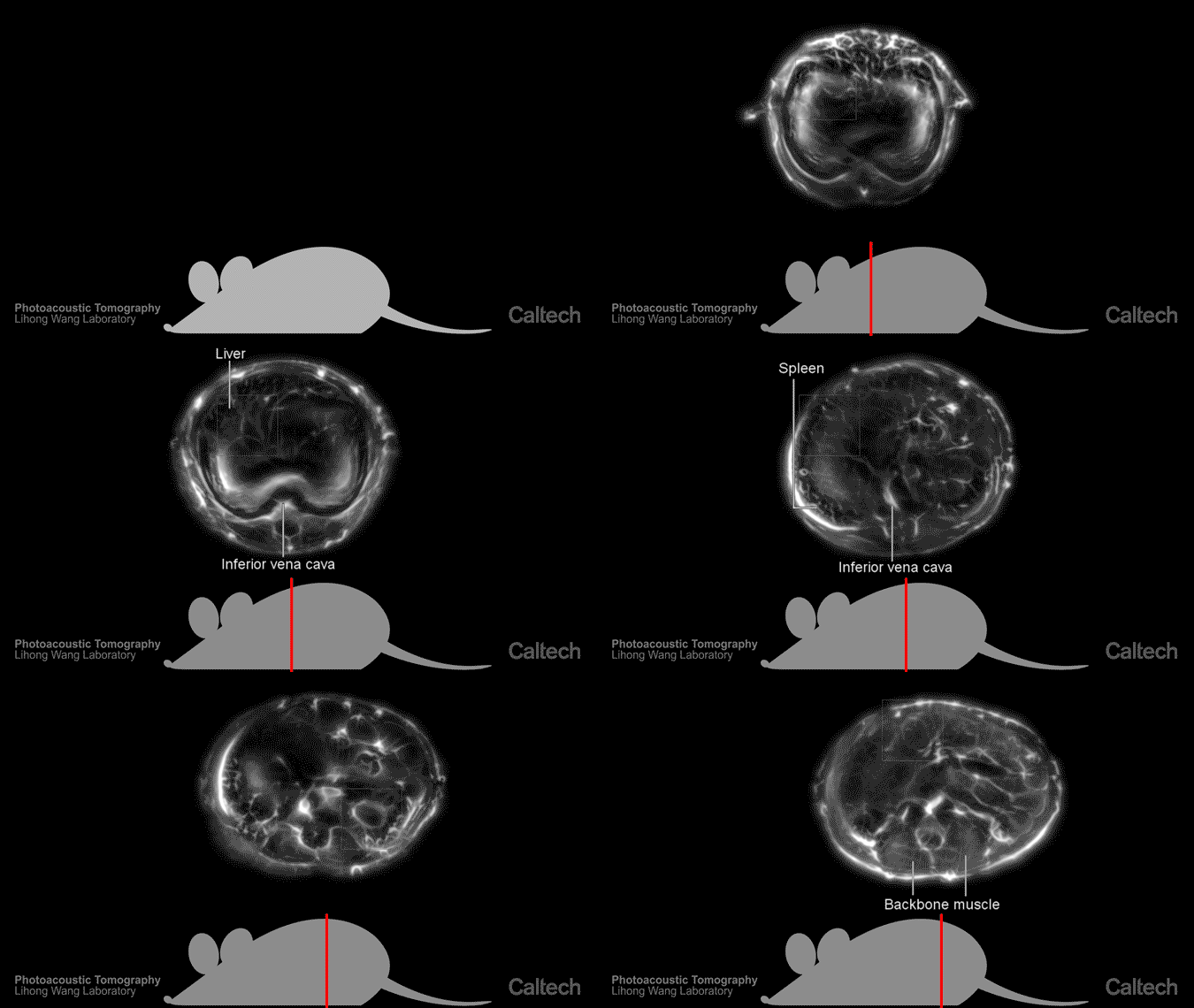
The picture shows an image of a mouse organ generated by photoacoustic imaging technology. This technology combines light with ultrasound to generate a complete tomogram in real time.


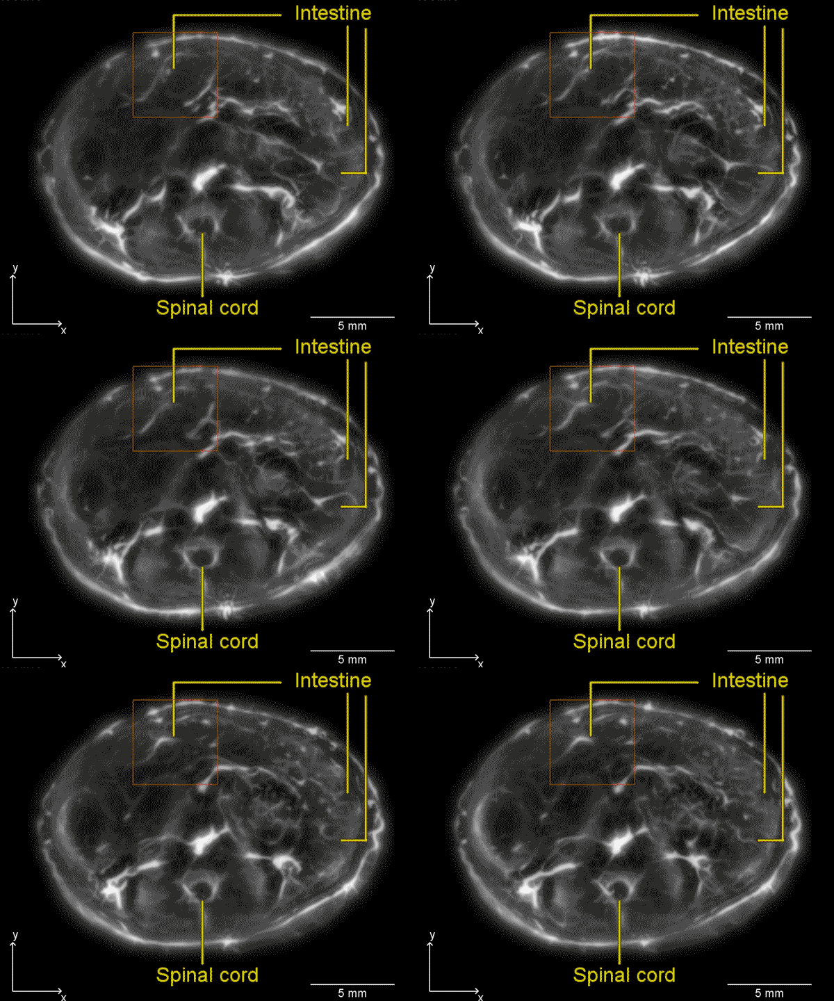
The picture shows the mouse gut image generated by photoacoustic imaging technology.
Sina Technology News Beijing time on May 25th news, according to foreign media reports, using a new laser imaging technology, scientists can view the internal conditions of small animals in real time. The technology is based on light and ultrasound, and the resolution is sufficient for scientists to see animal organs, flowing blood, diffuse melanoma cells, and efficient neural networks. In this way, researchers can monitor the diffusion of drugs in animals and understand how different organs respond to drugs.
The study was conducted jointly by Duke University and the California Institute of Technology. The technology is called "Single Pulse Photoacoustic Computed Tomography" (SIP-PACT for short), which uses light microscopy and ultrasound imaging to observe the animal's internal condition. Researchers say that in vivo scanning of small animals has always had limitations on image resolution and scanning speed, and this new technology can solve this problem.
It can generate tomograms in animals in real time. For example, in adult mice, 50 complete tomograms can be generated per second. "Photoacoustic imaging technology can generate complete tomographic images of small animal bodies in real time, and we have high hopes." Dr. Junjie Yao, co-author of the study and assistant professor of biomedical engineering at Duke University, pointed out that "using this technology, research Personnel can easily monitor the distribution of drugs in animals and the response of different organs to drugs."
Photoacoustic imaging technology integrates multiple imaging technologies on the same platform. Traditional light microscopy techniques can quickly generate high-resolution images that reflect the internal details of the animal's body through the wavelengths of light that are absorbed, reflected, or diverged by different tissues. For example, melanin absorbs near-infrared light, and blood's response to light depends on the amount of blood oxygen. However, since most of the light scatters as it penetrates the tissue, the imaging depth is only a few millimeters. In contrast, ultrasound can easily penetrate the body tissue, so we can observe more deeply. However, ultrasound cannot judge the chemical composition of the tissue, so it cannot provide us with important medical information like light.
Magnetic resonance imaging (MRI) can also observe the internal conditions of the tissue, but it requires a powerful magnetic field and generates images for a long time, ranging from a few seconds to a few minutes. X-ray and positron emission computed tomography (PET) It also generates a lot of radiation, which can't be used for long-term observation. However, photoacoustic imaging technology uses powerful and short-time laser pulses to make the cells emit ultrasonic waves and penetrate the body tissues under the premise of ensuring safety.
In this latest study, Dr. Yao and Dr. Lihong Wang of the California Institute of Technology have also significantly increased the speed and scanning range of photoacoustic imaging technology. They set up a circular ultrasound probe and a fast data acquisition system to determine the source of each ultrasound in small animals using triangulation. Finally, the improved imaging technique can penetrate the interior of the biological tissue by five centimeters. It is also in millimeters while retaining the information provided by traditional light microscopy.
“This is equivalent to compressing the sun collected in the afternoon of one afternoon into the size of a nail and then launching it in one nanosecond,†Dr. Yao explained. He has been working on this technology for nearly a decade. "After the laser hits the cell, the energy it carries causes the cell to heat up slightly and the volume expands immediately, which creates ultrasound, which is like the difference between slowly pushing an object and striking it and making it vibrate. We can understand the full range of information in the living body, and every laser pulse will not miss any details. It can monitor the dynamics of the organism in real time, such as the beating of the heart, the expansion of the artery, the function of different tissues, etc."
Using this technique, the researchers tracked the progression of melanoma cells in the blood vessels of mice, while taking pictures of the high-speed operation of the entire brain's neural network. “This technology is very powerful because it doesn't require any contrast agent to be injected.†Dr. Yao pointed out, “This way we can determine that changes in the body are not caused by external causes. We believe this technique is used in preclinical imaging and clinical diagnosis. The field has great potential." (Leaf)
Source: Sina Technology
Household Electric Appliances,Smart Home Appliances,Small Household Appliances
Ningbo Shuangtuo International Trade Co., Ltd. , https://www.nbst-sports.com